FAQ

Frequently Asked Questions

GLAUCOMA

What is glaucoma?

Glaucoma is a disease of the optic nerve that is associated with an elevated eye pressure, optic nerve changes, and visual field loss. When a patient has glaucoma, the fluid in the eye does not drain properly causing the pressure in the eye to increase. As the pressure increases, it pushes on the optic nerve causing damage. The damage caused by glaucoma cannot be reversed, however treatments are available to prevent any further vision loss. There are no symptoms of glaucoma, but the loss of peripheral vision can be detected with special testing. It is important to have your eye pressure checked and optic nerve examined yearly.

What causes glaucoma?

There are many causes of glaucoma but the common theme is an increased eye pressure that is associated with a poor drainage system in the eye. It can be associated with increasing age, cataracts, and medical problems like diabetes. There is a strong hereditary component as well.

Who is at risk for glaucoma?

High pressure alone does not mean that you have glaucoma. Your Ophthalmologist considers many factors in determining your risks for developing this disease. The most important risk factors include:

  • Age
  • Nearsightedness
  • African ancestry
  • A family history of glaucoma
  • Past injuries to the eyes
  • A history of severe anemia or shock

Your ophthalmologist will weigh all of these factors before deciding whether you need treatment for glaucoma or whether you should be monitored closely as a glaucoma suspect.This means your risk of developing glaucoma is higher than normal, and you need to have regular examinations to detect the early signs of damage to the optic nerve.

CATARACT

What is a cataract?

A cataract is a clouding of the normally clear lens of the eye. It can be compared to a window that is frosted or yellowed. There are many misconceptions about cataracts. A cataract is not:

  • A film over the eye
  • Caused by overusing the eyes
  • Spread from one eye to the other
  • A cause of irreversible blindness

Common symptoms of cataracts include:

  • A painless blurring of vision
  • Glare or light sensitivity
  • Poor night vision
  • Double vision in one eye
  • Needing brighter light to read
  • Fading or yellowing of colors

The amount and pattern of cloudiness within the lens can vary. If the cloudiness is not near the center of the lens, you may not be aware that a cataract is present.

How is a cataract treated?

Surgery is the only way a cataract can be removed. However, if symptoms of a cataract are not bothering you very much, surgery may not be needed. Sometimes a simple change in your eyeglass prescription may be helpful. There are no medications, dietary supplements, or exercises that have been shown to prevent or cure cataracts. Protection from excessive sunlight may help slow the progression of cataracts. Sunglasses that screen out ultraviolet (UV) light rays or regular eyeglasses with a clear, anti-UV coating offer this protection.

Will cataract surgery improve my vision?

The success rate of cataract surgery is excellent. Improved vision is achieved in the majority of patients if other vision-limiting problems are not present.

DIABETIC RETINOPATHY

What is diabetic eye disease?

Diabetic eye disease refers to a group of eye problems that people with diabetes may face as a complication of this disease. It is the leading cause of severe vision loss in the working population. It is more common in those with a long history of diabetes and in those patients that have had poor control of his/her blood sugars.

Diabetic eye disease may include:

  • Diabetic retinopathy – damage to the blood vessels in the retina.
  • Cataract – clouding of the eye’s lens.
  • Glaucoma – increase in fluid pressure inside the eye that leads to optic nerve damage and loss of vision.

Cataracts and glaucoma also affect many people who do not have diabetes.

What are the symptoms?

Often there are no symptoms in the early stages of the disease. Vision may not change until the disease becomes severe. Blurred vision may occur when the macula – the part of the retina that provides sharp, central vision – swells from the leaking fluid. This condition is called macular edema. If new vessels have grown on the surface of the retina, they can bleed into the eye, blocking vision. Unfortunately, advanced retinopathy can be present even without vision loss or symptoms and for that reason, an annual exam is recommended.

How is diabetic retinopathy treated?

The best treatment is strict control of your blood sugar. This alone will significantly reduce the long-term risk of vision loss from diabetic retinopathy. If high blood pressure and kidney problems are present, they need to be treated. Laser surgery has been the primary focus of therapy for many years but recently injection therapy has been shown to have superior visual results in some patients. These intraocular injections can cause regression of the hemorrhages, swelling and bleeding that are commonly associated with diabetic retinopathy. Laser still plays a role in treating retinopathy as does more complex vitrectomy surgery for traction retinal detachment and non-clearing vitreous hemorrhages.

What is macular degeneration?

Age related Macular degeneration (AMD) is a leading cause of vision loss in those over 65. The associated anxiety of losing vision and independence can best be addressed with an understanding of the person at risk, the symptoms, and the treatments that improve the prognosis if the diagnosis is made early.

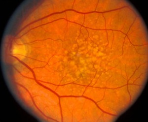
Macular degeneration is group of conditions that causes the central part of the retina, the macula, to lose the ability to see. This is an area behind the eye that cannot be easily seen or evaluated unless the eyes are dilated. There are two types of macular degeneration, wet and dry.

Dry macular degeneration is much more common and involves a slow process of aging cells that stop working and lead to blurred or missing areas in the central field of vision. It does not take away side vision.

Wet macular degeneration occurs when the aging retina grows new blood vessels that leak fluid and cause bleeding under the retina. These changes under the retina lead to sudden symptoms of blurred central vision and demand a prompt evaluation and treatment by an Optometrist.

It is much less common than dry AMD but receives more attention because of the potential for sudden severe loss of vision and because treatment is available. Patients are often ill prepared for the effect to lifestyle, hobbies, and work.

Macular degeneration helps improve the prognosis, but it is very important to understand that early macular degeneration often has no symptoms.Therefore, nothing replaces regular dilated eye exams and the American Academy of Ophthalmology suggests an exam every one to two years after the age of 65.

Patients must remain vigilant by testing each eye alone for more advanced symptoms: blurred vision, distortion, or missing areas in the central vision. The patient may notice it is more difficult to see faces or read, but it is important to remember that with both eyes open, advancing macular degeneration in one eye can be missed.

One can avoid this by checking vision with one eye covered while reading the paper or doing a Sudoku puzzle. Symptoms such as bent/curved lines, or missing letters can be noticed with single corrective eye exam.

Monitoring your vision is good at all ages but from a practical standpoint we can use our personal risk factors to guide our frequency of monitoring for symptoms. While we recognize an increased risk in blue eyed Caucasians of northern European descent, there is three risk factors that demand greater attention: a positive family history, a history of past or present smoking, and advancing age. Every race has an increased risk with advancing age, but a family history can be the most predictive. Genetic testing is not recommended yet but patient with a positive family history of AMD requires earlier and more frequent exams to identify early disease and to allay unnecessary fears if the exam is normal.

While you can’t pick your genetics or stop aging, smoking is the most important risk factor that you can modify. If you are concerned about vision loss from AMD, you must stop smoking. After your dilated macular exam, you and your doctor can tailor a reasonable frequency for exams and home monitoring for advancing AMD.

MACULAR DEGENERATION

How is macular degeneration diagnosed?

If monitoring detects new distortion, blurred vision, or areas of missing vision, you should seek prompt consultation with your eye doctor. Early diagnosis is important because it improves prognosis. Diagnostic evaluations may be as simple as a dilated retinal exam with your doctor but could also include an optical coherence tomography (OCT).

This is a retinal scan that can detect small amounts of retinal fluid that may otherwise be missed. More advanced testing with a retina specialist may include an injection of a contrast agent into your arm to perform an angiogram of the retinal vessels to look for abnormal new vessels. These tests determine your potential need for treatment.

Treatment of wet AMD has seen great strides over the past 10 years. The prognosis has greatly improved with the introduction of injectable medication. Patients who were once certain to become blind have a much better chance of maintaining useful vision.

The importance of early treatment is highlighted by the 90% of people who will maintain or have improved vision if injection treatment is started during early onset of the disease. Regular injections of Eylea, Lucentis, or off- label use of compounded Avastin suppresses leakage and bleeding but unfortunately does not cure the disease. While a few patients can stop therapy, most require regular monitoring and injections indefinitely.

There is no current treatment for dry macular degeneration, but research is looking for ways to stop the degenerative process. Most therapy is supportive in nature with low vision aids to help with reading and daily tasks. Low vision aids can also be of great help to patients with wet AMD.

Vitamin therapy is based on the Age Related Eye Disease Studies that evaluated the role of high dose vitamin therapy at reducing the incidence of AMD and vision loss. In the AREDS II trial, those patients taking the supplements with lutein/zeaxanthin had a 26% reduced risk of developing advanced AMD compared to those not taking the supplement. Most of the participants also took a multivitamin.

Macular degeneration is a complex topic that can cause anxiety because it is poorly understood, and we all fear the loss of vision and independence. With education, routine monitoring, and prompt therapy, the poor vision, and fear of AMD can be minimized.

We stress regular dilated eye exams, not smoking, and a diet in green leafy vegetables and AREDS II vitamin supplements to reduce its effect.

Screening:

To detect AMD before symptoms appear, you should be screened if you meet any of the following criteria:

  • Over the age of 60
  • Have hypertension or cardiovascular disease
  • Smoke cigarettes or other tobacco products
  • Have a close relative (sibling, parent, or child) with AMD related vision loss
  • Had cataract surgery (replacement of the crystalline lens of the eye with an artificial lens)
  • Had significant cumulative light exposure (e.g., airline pilots, boaters, farmers)

Understanding Your Diagnosis:

Early detection of AMD is essential to preserving your vision. That’s why it’s important to learn everything you can about AMD. Arrive for your appointments prepared with notes on your symptoms and ask questions about anything you don’t understand. Take an active role in preventing vision loss.